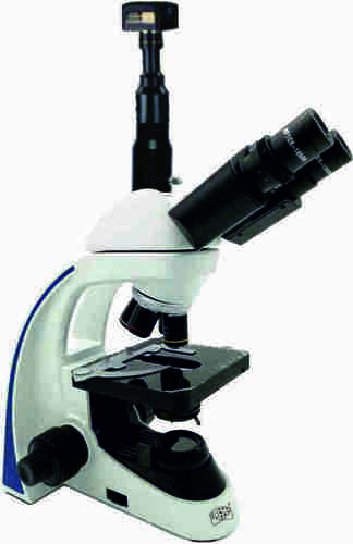
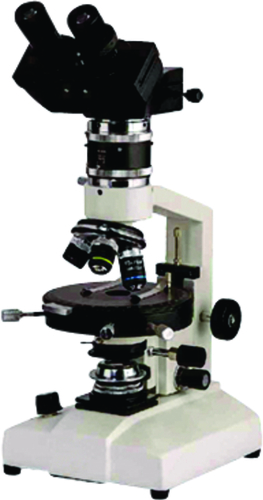
About Research Trinocular Microscope with camera model medi-vision
Body Ergonomic designed SingleMold Low Drive Coaxialmechanismfor comfortable basedonballbearingandwireguides
Viewing Bodies SideNTopButter y type 30 inclined Trinocular observation HeadIncorporatedwith anti fungus coated anti reection coated Prisms Rota table to 360 withinter pupillarydistance4875mm5diopteradjustablewithlockingdevice
NosepieceQuadrupleforcenteredalignmentofobjectives
FocusingCoaxial Coarse ne Movement slides smoothly on Ball Bearing Guide Ways which providesthehighestDegreeofsensitivemorethantwomicronsnefocusingreadingto0002freefrombacklashMechanicalStage Low driveHorizontal coaxial mechanical stage working on Ball Bearing Guide Waysof145x140mmwithneVeniregraduationsreading01mmXYmovement75mmx55mmofslides
CondenserPre centered Abbecondenser NA 125 with aspheric lens with Iris diaphragm Built in swingoutlterholderwithlterupdownmovementthroughrackpiniononstainlesssteelguides
IlluminationHeavy rectangular sturdybase with built in LED Illumination Batterybackupoptionalorhalogenlamponoffswitchcontinuouslyvariablelightintensitycontrolledregulatorquickchangebulbprovisionworkableon110240v50HzAC
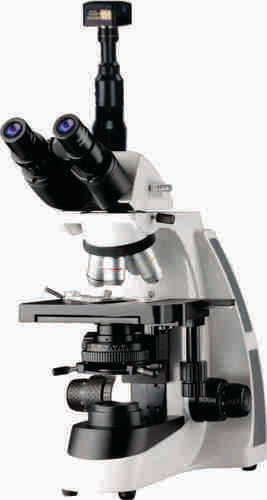
Advanced Optical Performance for Research ExcellenceThis model incorporates plan achromatic objectives and a bright field illumination system, ensuring sharp, true-color images essential for critical laboratory work. Its anti-fungal optical coating preserves image clarity over time, making it a reliable partner in demanding research environments, clinical laboratories, and academic settings.
Ergonomic and Versatile DesignFeaturing a 45 inclined, 360 rotatable trinocular head, and wide interpupillary range, the microscopes modular design makes it comfortable for extended observation sessions. Coarse and fine coaxial focusing, alongside a precision stage, provide smooth sample navigationdelivering accuracy during high-magnification studies.
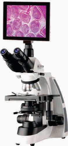
Comprehensive Digital Imaging SuiteThe integrated 5.0 MP digital camera, high frame rate video, and Windows-compatible analysis software enable real-time observation and thorough documentation of samples. Image capture in multiple formats (JPEG, BMP, TIFF, PNG) simplifies data sharing and archiving for diagnostics, research, and educational presentations.
FAQs of Research Trinocular Microscope with camera model medi-vision:
Q: How can I capture and analyze images using the included camera software with the medi-vision trinocular microscope?
A: Simply connect the integrated digital camera to your PC using the USB 2.0 interface. Install the provided driver and image analysis suite (compatible with Windows). This lets you view live images, capture still photos at up to 5.0 MP resolution, record videos, and analyze data in multiple file formats such as JPEG, TIFF, BMP, or PNG.
Q: What benefits does the anti-fungal optical coating provide?
A: The optics are coated with an anti-fungal treatment, protecting lenses and optical surfaces from fungal growth in humid or laboratory environments. This ensures lasting image clarity, reduces maintenance, and extends the life of your microscopes high-performance optics.
Q: When is it ideal to use the medi-vision microscopes different color filters?
A: The swing-out filter holder includes blue, green, and yellow filters that enhance specimen contrast for various sample types. For instance, blue filters improve resolution and contrast under bright field illumination, while green filters assist when examining stained biological tissues, ensuring optimized visualization.
Q: Where can the medi-vision research microscope be effectively used?
A: It is perfectly suited for research laboratories, pathology, biological sciences, clinical diagnostics, and educational institutions in India and globally. Its robust build, precision optics, and digital imaging support diverse applications, from research analysis to routine classroom demonstrations.
Q: What makes the focusing and stage movement precise and user-friendly?
A: This microscope features coaxial coarse and fine focus controls with long travel and tension adjustment, allowing precise focusing to within 0.002 mm. Its double-layer mechanical stage with rack and pinion X-Y translation provides accurate and smooth sample positioning, supporting detailed examination at high magnifications.
Q: How does the microscope ensure true color representation of specimens?
A: With integrated true color digital image processing in the camera and plan achromatic objectives, the microscope accurately reproduces the natural hues of specimens, which is critical for diagnostic and research accuracy. Users can reliably document and analyze their findings with minimal color distortion.


 Send Inquiry

