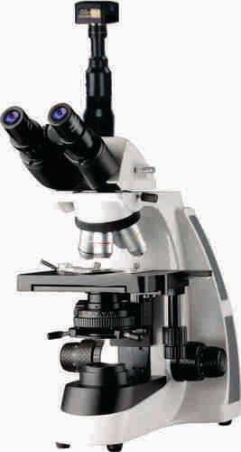
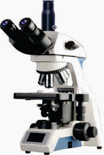
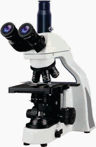
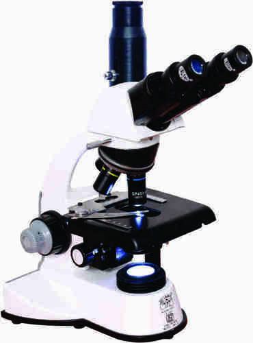
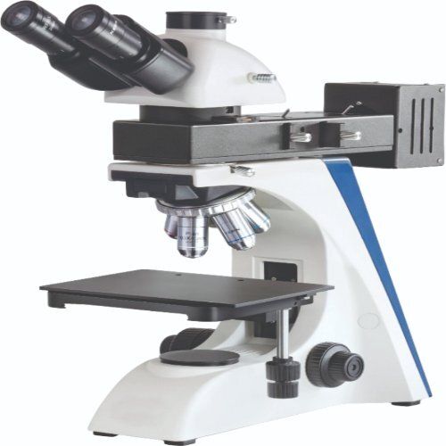
About Research Trinocular Microscope with Camera Model True-Vision
Body Ergonomic designed SingleMold Low Drive Coaxialmechanismfor comfortable basedonballbearingandwireguides
Viewing Bodies SideNTopButter y type 30 inclined Trinocular observation HeadIncorporatedwith anti fungus coated anti reection coated Prisms Rota table to 360 withinter pupillarydistance4875mm5diopteradjustablewithlockingdevice
NosepieceQuadrupleforcenteredalignmentofobjectives
FocusingCoaxial Coarse ne Movement slides smoothly on Ball Bearing Guide Ways which providesthehighestDegreeofsensitivemorethantwomicronsnefocusingreadingto0002freefrombacklashMechanicalStage Low driveHorizontal coaxial mechanical stage working on Ball Bearing Guide Waysof145x140mmwithneVeniregraduationsreading01mmXYmovement75mmx55mmofslides
CondenserPre centered Abbecondenser NA 125 with aspheric lens with Iris diaphragm Built in swingoutlterholderwithlterupdownmovementthroughrackpiniononstainlesssteelguides
IlluminationHeavy rectangular sturdybase with built in LED Illumination Batterybackupoptionalorhalogenlamponoffswitchcontinuouslyvariablelightintensitycontrolledregulatorquickchangebulbprovisionworkableon110240v50HzAC
Advanced Optical and Digital PerformanceEnjoy high-contrast images and precise measurements with achromatic objectives, a quadruple revolving nosepiece, and a high-resolution camera. The versatile magnification range and ergonomic design ensure suitability for demanding research and clinical environments.
Seamless Camera Integration and Software SupportThe microscope's standard C-mount and USB-powered 5MP camera allow effortless digital imaging. Included software delivers robust editing, measurement, and annotation tools-and is compatible with Windows 7/8/10/11 and Mac OS-making documentation and analysis straightforward.
Durability and Usability for Professional SettingsConstructed with a sturdy, powder-coated metal frame, the True-Vision microscope endures daily laboratory use. Its foam-fitted export packaging and supplied accessories (dust cover, immersion oil, cleaning kit, power cord) make it ideal for busy research, university, and clinical settings.
FAQ's of Research Trinocular Microscope with Camera Model True-Vision:
Q: How do I connect the digital camera to my computer and operate it?
A: Simply attach the camera to the microscope's C-mount and connect it to your computer via USB. The camera is powered through this connection. The included software supports both Windows (7/8/10/11) and Mac OS, enabling easy image and video capture, editing, measurement, and annotation.
Q: What types of applications is the True-Vision Trinocular Microscope best suited for?
A: This microscope is ideal for research laboratories, universities, pathology, and clinical diagnostics, offering precise imaging and robust digital documentation. It is also suitable for advanced teaching and routine laboratory analysis where high-resolution imaging is essential.
Q: What is the process for changing objectives and adjusting focus?
A: Switch between the four achromatic, spring-loaded objectives using the ball-bearing mounted quadruple revolving nosepiece. Coarse and fine coaxial focus knobs provide smooth, precise focusing, with a fine adjustment graduation of 0.002 mm and coarse adjustment up to 30 mm.
Q: Where can I use this microscope regarding power and portability?
A: The microscope features a switchable power supply (AC 220V/50Hz or AC 110V/60Hz), making it compatible with international voltage standards. Its sturdy build and foam-fitted export packaging facilitate safe transport and installation in various laboratory environments.
Q: What are the benefits of the included imaging software?
A: The software provides comprehensive tools for capturing images and video, editing, measurement, and annotation. This streamlines data analysis, documentation, and sharing in both research and diagnostic workflows.
Q: When should users utilize the oil immersion lens, and how is it maintained?
A: Use the 100x Oil (spring loaded) achromatic objective for observing fine cellular details, especially in research and pathology applications. Maintenance is straightforward-use the supplied immersion oil sparingly and clean the lens after each use with the provided cleaning kit.
Q: What formats and resolutions are supported for image and video capture?
A: The camera captures still images up to 2592 x 1944 pixels and records video at 1920 x 1080 pixels, with a frame rate of 30 fps at lower resolution. Supported image formats include JPEG, BMP, TIFF, and RAW, facilitating flexible image management for publication and analysis.


 Send Inquiry

