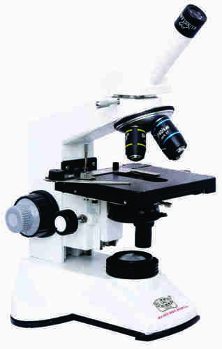
Pathological monocular Microscope Medilux -m About
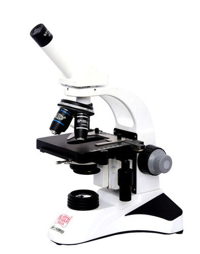
Ergonomic designed for comfortableprolonged microscopy, highly rigid, strong, easy to carry,ViewingBodies: - Monocular, Binocular or Trinocular (any one) observation Head inclined atNosepiece:Quadruple,forcenteredalignmentofobjectives.
Focusing:-Coaxial Coarse &ine Movement slides smoothly on Ball Bearing Guide Ways which providesthehighestDegreeofsensitivemorethantwomicronsinefocusingreadingto0.002freefrombacklash,MechanicalStage:Condenser:-PrecenteredAbbecondenserNA1.25withasphericlenswithIrisdiaphragmBuilt-inswingoutilterholder,up-downmovementthroughrack&piniononstainlesssteelguides.
Illumination:-Heavy rectangularsturdy base with built in LED Illumination (Battery backupoptional)orhalogenlamp,on/offswitch,continuouslyvariablelightintensitycontrolledregulator&quickchangebulbprovisionworkableon110-240v,50HzAC.
Packing:-completewithduster,cover,Necessarycords&cables&instructionmanual.
Achieve Superior Clarity with Achromatic ObjectivesEquipped with achromatic objectives (4x, 10x, 40x, 100x), the Medilux-m delivers distortion-free images across a wide magnification range. Users experience enhanced color correction and sharp focus, ideal for thorough pathological analyses. These lenses are perfect for differentiating intricate cellular structures and provide reliable results in clinical and research settings.
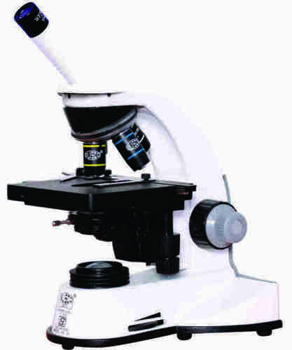
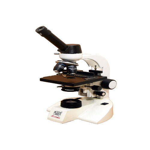
User-Friendly Monocular DesignThe Medilux-m features a single drawtube for comfortable monocular viewing. Its standard 10x widefield eyepiece and simple, sturdy construction make it exceptionally user-friendly. The adjustable viewing head helps users achieve optimal viewing angles and minimizes eye fatigue during prolonged use.
Precision Focus and IlluminationCoarse and fine adjustment knobs allow precise focusing, essential for observing delicate specimen details. The built-in LED light source provides consistent illumination, while the condenser lens enhances the intensity and contrast of the field of view. Together, these features ensure a bright and clear image for detailed examination.
FAQ's of Pathological monocular Microscope Medilux -m:
Q: How do I use the Medilux-m monocular microscope for pathological examinations?
A: Begin by placing your specimen slide securely on the two-layer mechanical stage with slide clips. Use the coarse adjustment knob to locate your sample with low magnification, then refine the image with the fine adjustment knob. Select the appropriate achromatic objective (4x, 10x, 40x, 100x) depending on the detail you wish to observe.
Q: What are the key features that distinguish the Medilux-m microscope?
A: The Medilux-m features a single monocular drawtube, a standard 10x widefield eyepiece, achromatic objectives (4x, 10x, 40x, 100x) providing 40x-1000x magnification, a built-in LED light source, and a mechanical two-layer stage. These components combine to deliver high-resolution, sharp, and color-accurate images.
Q: When should I use coarse versus fine adjustment for focusing?
A: Use the coarse adjustment knob when first locating your specimen, especially under lower magnifications. For higher objectives and detailed viewing, switch to the fine adjustment knob to achieve sharp, precise focus without disturbing your sample.
Q: Where is the LED illumination system located, and how does it enhance viewing?
A: The LED illumination system is integrated into the microscope's base, providing a stable and bright light source directly beneath the stage. This setup produces even illumination, enhancing visibility and contrast for clear observation of stained and unstained specimens.
Q: What is the advantage of using achromatic objectives in this microscope?
A: Achromatic objectives minimize optical distortions, ensuring clear, true-to-color images across the microscope's magnification range. This is particularly beneficial in pathology, where accurate color representation and detail are crucial for analysis.
Q: How does the two-layer mechanical stage benefit my workflow?
A: The two-layer mechanical stage allows smooth and precise positioning of slides using adjustment controls. This feature facilitates systematic scanning of specimens, making it easier to locate and examine desired areas efficiently during continuous observation.






 Send Inquiry


 Send Inquiry