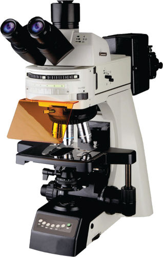
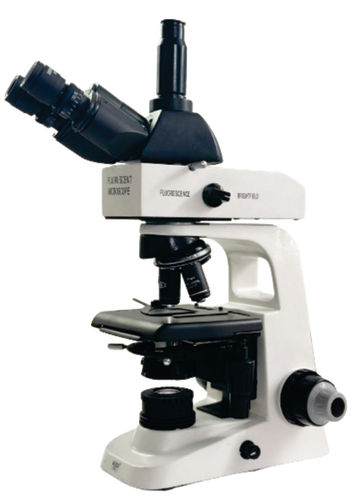
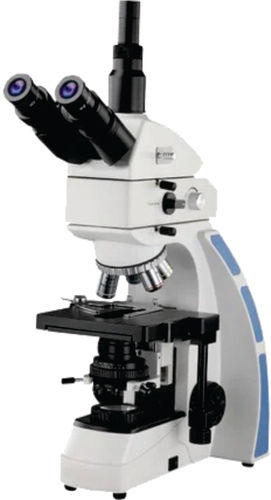
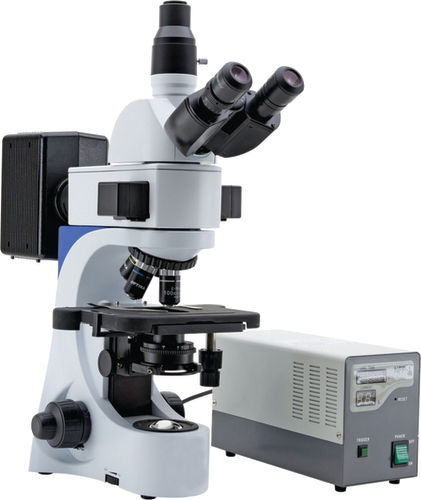
Research Fluorescent Microscope 6 Filters Model FL-600 About
Eyepiece
Wideeldeyepiece10XpairedFOV25mm
ObjectivesAntifunguscoated
Objective10X
MechanicalBody
anglesixtuplenosepieceonballbearingSlotforPolarizerandDIC
SubstageCondenser
SwingoutfrontlensNA09125lterholderandirisdiaphragm
ExcitationFiltersOptional
Systemwith6positionreectorturretDAPIFITCTRITCCY5capableofcapturinganydye
LightSource
FL600
StandardAccessories
3W LED 12v100wHalogen bulb operating manual dust cover fuse guarantee card blue lterpower supply for100wmercurylampandstyrofoammoldedpack
Innovative Fluorescence ImagingThe FL-600 microscope utilizes LED fluorescence illumination and six integrated filters, enabling users to investigate a variety of fluorescently labeled specimens with outstanding clarity. Its plan achromatic objectives provide sharp images across the field of view, supporting advanced research in cell biology, microbiology, and material science.
Precision and Comfort CombinedDesigned with ergonomics in mind, the binocular head is inclined at 30, suited for extended observation sessions. The wide 55-75 mm interpupillary range accommodates various users, while the double-layer mechanical stage and fine focus controls allow for precise manipulation and focusing of samples.
Seamless Digital DocumentationWith an advanced CMOS sensor and USB 3.0 connectivity, the FL-600 enables instant capture of still images and videos at 1920 x 1080 resolution. The microscope supports both JPEG and BMP formats, making data archiving and sharing efficient and straightforward for professionals and educators alike.
FAQ's of Research Fluorescent Microscope 6 Filters Model FL-600:
Q: How does the built-in filter system enhance fluorescent imaging in the FL-600 microscope?
A: The FL-600 includes six integrated filters, which enable selective excitation and emission of specific fluorophores. This capability allows researchers to visualize multiple fluorescent labels with precision, producing clear and distinct images for multi-channel fluorescence applications.
Q: What types of specimens can be observed using the FL-600 research fluorescent microscope?
A: This microscope is designed to examine a variety of samples, particularly those labeled with fluorescent dyes such as biological cells, tissues, and microorganisms. It is suitable for research in areas like molecular biology, medical diagnostics, and materials science.
Q: When is digital imaging with the FL-600 especially beneficial?
A: Digital imaging is crucial during documentation, analysis, and sharing of visual data with colleagues or students. The FL-600's high-definition sensor and 30 fps frame rate enable real-time viewing and recording, supporting research presentations and publication needs.
Q: Where can the FL-600 microscope be effectively utilized?
A: The FL-600 is well-suited for use in research laboratories, educational institutions, clinical diagnostics, and industrial quality control. Its robust features accommodate applications requiring advanced fluorescence and precise image capture.
Q: What is the process for capturing and saving images with this microscope?
A: Users can capture still images or record videos directly through the CMOS sensor. Data is transferred via the USB 3.0 interface to a connected computer, where images can be saved in JPEG or BMP format at a resolution of 1920 x 1080 pixels.
Q: How does the adjustable interpupillary distance benefit users of the FL-600?
A: The adjustable interpupillary distance (55-75 mm) allows users to set the eyepiece spacing for optimal comfort and clear binocular vision, enhancing both accuracy and ease during extended observation periods.
Q: What are the primary advantages of using the FL-600 research fluorescent microscope?
A: Key benefits include high-resolution fluorescence imaging, ergonomic design, precise mechanical stage control, and seamless digital documentation. These features together elevate the accuracy and efficiency of advanced scientific research and analysis.






 Send Inquiry


 Send Inquiry