Research Fluorescent Microscope 4 Filters Model Fl-400 About
Eyepiece
Extra wideeld10XpairedFOV22mm
MechanicalBody
angle sixtuplenosepiece onball bearingSlotforPolarizer andDIC
division0001mmcorseperrotationnestroke01mmper rotationmovingrange
SubstageCondenser
SwingoutfrontlensNA09125lterholderandirisdiaphragm
FL400
ExcitationFiltersOptional
LightSource
TransmittedKohlerlightsource3WLEDor12V100Wtungstenhalogenbulb
EpiFluorescencelightsource100Whighpressuremercurylamp
High-Resolution Research ImagingBenefit from sharp, vivid imaging with the FL-400's 5-megapixel CMOS sensor. Capture still images at 2560 x 1920 pixels and HD video at 1920 x 1080 pixels, ensuring every detail in fluorescence is preserved for analysis. The USB 3.0 interface streamlines transferring data for quick reporting and documentation.
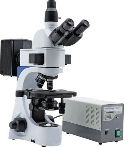
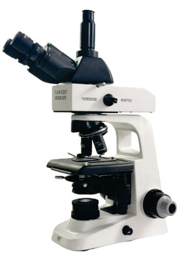
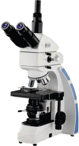
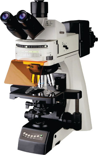
Precision and Comfort for ResearchersDesigned with a Seidentopf binocular head, inclined at 30 degrees, the FL-400 ensures ergonomic comfort during extended observation sessions. Its double-layer mechanical stage with X-Y movement and fine adjustment (0.002 mm) delivers ease and control for intricate research. The widefield WF10X/18 mm eyepieces and adjustable interpupillary range accommodate diverse users.
Advanced Fluorescence CapabilitiesUtilizing four filters and a bright yet adjustable 3W LED light source, the FL-400 amplifies fluorescence signals for detailed cellular studies. The Abbe condenser with iris diaphragm and achromatic objectives (4X, 10X, 40X, 100X) provide excellent clarity and magnification for cutting-edge research applications.
FAQ's of Research Fluorescent Microscope 4 Filters Model Fl-400:
Q: How do I capture high-resolution still images and videos with the FL-400 microscope?
A: Connect the FL-400 microscope to your computer via USB 3.0. Still images can be captured at a resolution of 2560 x 1920 pixels, while video recording is available at 1920 x 1080 pixels and 60 fps. Files are saved in JPEG and BMP formats for convenient analysis and sharing.
Q: What types of research benefits from using this fluorescent microscope model?
A: The FL-400 is ideal for cellular, molecular, and fluorescence-based research in biology, medical, and materials science fields. Its advanced optics, four fluorescence filters, and precise focusing mechanisms make it well-suited for observing details in stained or fluorescently-tagged specimens.
Q: Where can the FL-400 microscope be installed and used effectively?
A: This microscope is perfect for laboratories, research institutions, universities, and clinical settings across India. Its compact dimensions (400 mm x 250 mm x 450 mm) and rotatable binocular tube make it suitable for both individual workstations and shared research environments.
Q: What advantage does the adjustable LED illumination provide?
A: The LED illumination, with adjustable brightness, ensures consistent lighting for various specimen types and minimizes heat exposure, which helps preserve sensitive samples during lengthy observations.
Q: How does the double-layer mechanical stage aid the research process?
A: The 140 mm x 140 mm double-layer mechanical stage, featuring X-Y movement, allows precise positioning and smooth navigation of slides. This ensures repeatable, accurate observations, especially when examining multiple loci or conducting comparative studies.
Q: What is the process for changing objective lenses on the FL-400?
A: The FL-400 is equipped with a revolving nosepiece that holds achromatic objectives (4X, 10X, 40X, 100X). Simply rotate the nosepiece to switch between magnifications, ensuring the correct lens is positioned and secured for your research needs.






 Send Inquiry


 Send Inquiry